Why glaucoma screening matters
Glaucoma is a group of conditions that damage the optic nerve, usually — but not always — associated with elevated pressure inside the eye. It is one of the leading causes of irreversible blindness worldwide, and the most common form, open-angle glaucoma, develops without pain, redness, or noticeable blur until vision loss is already advanced.
By the time a patient notices a problem, significant optic nerve damage has often already occurred and cannot be undone. The damage that comes before symptoms is what an eye exam is designed to catch.
What a comprehensive eye exam includes for glaucoma
A comprehensive eye exam that takes glaucoma seriously assesses both the structure of the optic nerve and the function of the visual pathway. The core elements:
- Tonometry — measurement of intraocular pressure. Elevated pressure is the most well-known risk factor for glaucoma, though glaucoma can occur at normal pressures too.
- Optic nerve examination — a direct view of the optic disc, usually with a dilated pupil, looking for changes in cup-to-disc ratio, asymmetry between the two eyes, disc haemorrhages, and thinning of the neuroretinal rim.
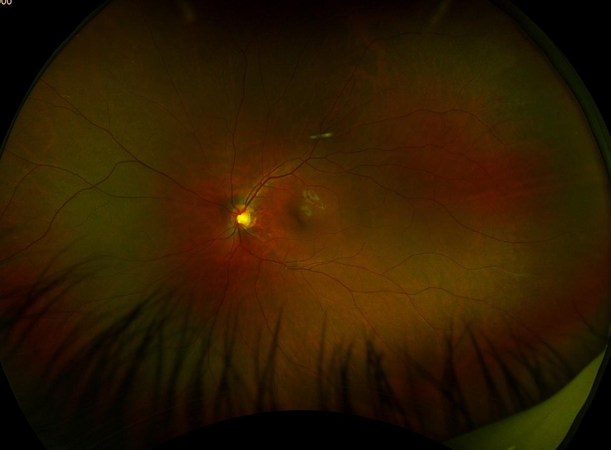
- Retinal photography — high-resolution images of the optic nerve and surrounding retina, stored and compared visit to visit. Subtle changes between visits are often the earliest sign of progression.
- OCT (optical coherence tomography) — cross-sectional scans that measure the thickness of the retinal nerve fibre layer and ganglion cell complex around the optic nerve. OCT can detect structural loss before it shows on a visual field test.
- Visual field testing — a functional test that maps where the patient can and cannot see, identifying areas of peripheral vision loss caused by nerve fibre damage.
- Pachymetry — corneal thickness measurement, which affects how pressure readings are interpreted. Thin corneas are an independent risk factor.
Not every patient needs every test at every visit. A baseline comprehensive screen is part of routine care; advanced testing is added when clinical findings, risk factors, or a suspicious optic disc make it appropriate.
What the optic nerve tells us
The optic nerve is the only part of the central nervous system that can be seen directly without surgery. Photographing it well — and comparing photos year over year — is one of the most clinically useful things a comprehensive eye exam does.
Who is at higher risk
Glaucoma can affect anyone, but several factors meaningfully raise risk:
- Age over 60 — risk rises steeply with age
- Family history — a parent or sibling with glaucoma is a strong risk factor
- African or Hispanic descent — earlier onset and faster progression on average
- Elevated intraocular pressure — the single biggest modifiable risk factor
- High myopia — longer eye shape changes the optic nerve and increases risk
- Diabetes
- Long-term steroid use — including topical drops, inhalers, and oral steroids
- Previous eye injury or surgery
- Thin corneas
Patients with multiple risk factors benefit from more frequent monitoring and from adding OCT and visual field testing earlier than a routine schedule would suggest.
What detection looks like before symptoms
Open-angle glaucoma damages peripheral vision first. The brain fills in missing areas, and the patient typically does not notice anything is wrong until central or near-central vision is affected — at which point a significant fraction of optic nerve fibres have already been lost.
The order of detection in a screening exam is usually:
- A structural finding first — a suspicious optic disc on examination or photography, or thinning of the retinal nerve fibre layer on OCT
- A functional finding next — a defect on visual field testing
- Symptoms last — and by this point, treatment shifts to slowing further loss rather than recovering what is already gone
This is why baseline imaging matters. A single exam tells you what the optic nerve looks like today. A second exam two years later tells you whether anything is changing — and that comparison is what catches early disease.
OHIP coverage for glaucoma in Ontario
OHIP covers comprehensive eye exams for:
- Children under 20 — annually
- Adults 65 and older — every 18 months
- Adults 20 to 64 with qualifying conditions, which includes glaucoma requiring treatment with medication, laser, or surgery
If a patient is being monitored as a “glaucoma suspect” but does not yet require treatment, OHIP coverage is generally not available — most working-age adults pay out of pocket or through private insurance until a treatment threshold is reached.
OHIP covers the base exam. Advanced imaging — OCT of the optic nerve, visual field testing, and supplementary retinal imaging — is beyond what OHIP funds and involves a supplemental fee. This imaging is often reimbursable through private insurance.
For full details, see OHIP eligibility and eye exam coverage in Ontario.
How often should I be screened?
A general guide:
- No risk factors, adult: a comprehensive exam every one to two years includes glaucoma screening as part of routine care
- One or more risk factors: annual exams, with OCT and visual fields added when clinically appropriate
- Glaucoma suspect: typically every six to twelve months, with imaging and field testing on a defined schedule
- Diagnosed glaucoma, under treatment: every three to six months, with monitoring coordinated alongside an ophthalmologist or glaucoma specialist
Recall intervals should be set by what the optometrist finds, not by an insurance cycle.
What to look for when comparing Toronto clinics
If you are choosing a clinic specifically with glaucoma screening in mind, useful signals include:
- The clinic measures eye pressure at every exam, not occasionally
- Optic nerve imaging is part of the standard comprehensive exam, not an upsell that depends on the day
- OCT and visual field testing are available on-site (faster, easier follow-up) rather than only by referral elsewhere
- Findings are tracked over time with a system that lets the optometrist compare year over year
- The optometrist will show you your own images and explain what is being watched
- There is a clear process for coordinating with an ophthalmologist when treatment is needed
What we do at Spadina Optometry
Every comprehensive eye exam at Spadina Optometry includes intraocular pressure measurement, direct examination of the optic nerve, and retinal photography that documents the optic disc for visit-to-visit comparison. When findings or risk factors call for it, we add OCT scanning of the retinal nerve fibre layer and visual field testing on-site.
Our optometrists explain findings in plain language, show patients their own optic nerve images, and coordinate referral to a glaucoma specialist when treatment is indicated. For patients already diagnosed with glaucoma or being co-managed with a specialist, we set monitoring intervals based on clinical findings and share imaging and reports with the rest of the care team.
If you have a family history of glaucoma, are over 60, or have not had a comprehensive eye exam in the last two years, glaucoma screening is one of the most clinically valuable reasons to come in.
Related
Want a comprehensive exam that takes glaucoma seriously?
We screen every patient and adjust the depth of testing based on age, family history, and clinical findings.
Prefer to talk first? Call or text us at 416-703-2797.
Spadina Optometry has cared for downtown Toronto patients since 2002 — an independent practice where the same team knows your eyes year after year.
Last reviewed: June 17, 2026